From the article you can find out what the parathyroid gland is, how, when and from what this organ is formed, what tasks it is designed to solve and where it is located. In addition, there is an informative video in this article. Data on the work of glandulae parathyroideae and their internal structure supplemented with photo materials from which you can find out how many parathyroid glands exist in human body and how they are arranged.
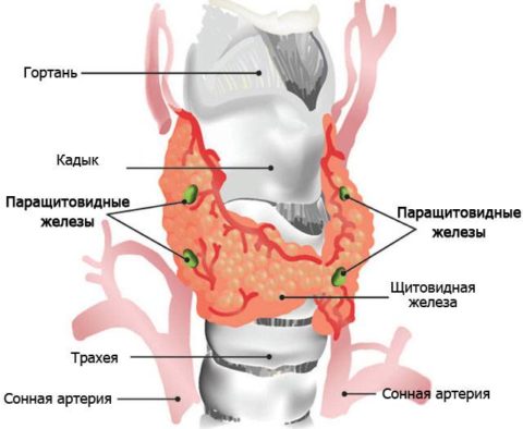
The parathyroid glands are small bodies (0.4-0.8 cm long, 0.3-0.4 cm wide and 0.15-0.3 cm thick), which are located on the posterior surfaces of both lateral lobes of the thyroid gland.
At first glance, these formations may resemble a variety of anatomical structures:
- Fat lobes.
- accessory thyroid glands.
- Split off parts of the thymus gland.
The thyroid and parathyroid glands differ from each other in the color of the substance, the latter are lighter, pale pink in color. childhood and yellowish in adults.
Variations in the number and location of glandulae parathyroideae
The development of the parathyroid gland occurs in the embryonic period from such anatomical structures as III and IV gill pockets. Their number at various people can be quite different. The frequency of occurrence of one or another number of glandulae parathyroideae is shown in the table:
These glands in some cases are completely immersed in the thyroid tissue, but most often located on its surface. The average weight of each of them is 0.05 - 0.09 g, and the total weight, as a rule, does not exceed 1.2 g.
Nerves and vessels glandulae parathyroideae
The supply of these organs of internal secretion with blood is carried out through the branches of the following arteries:
- Thyroidea superior.
- Thyroidea inferior.
- Arteries of the trachea.
- Arteries of the esophagus.
Blood from the arteries passes through wide sinusoidal capillaries and is collected in the following veins:
- Thyroideae inferiores.
- Thyroideae superiores.
- Plexus thyroideus impar.
The first two vessels form a plexus, and the third anastomoses with the pharyngeal and laryngeal veins.
Each artery supplying the parathyroid glands branches into an extensive network of capillaries that surround the parathyroid cells from different sides. Further, all the capillaries converge into a weaving network of veins, anastomizing with each other, which are collected in larger formations - venous subcapsular plexuses connected to the venous apparatus of the thyroid gland.
The innervation of the parathyroid glands is carried out from the same sources as the thyroid glands:
- laryngei inferiores (nn. Vagi).
- laryngei superiores (nn. Vagi).
- rr. sympathici (truncus sympathicus).
At the same time, the network of nerve endings is very saturated.
Anatomy of glandulae parathyroideae
Glandulae parathyroideae are found on both lateral lobes of the thyroid gland, and in some cases they are all localized on one side. The parathyroid glands are immersed in loose fiber that fills the space between the fascial sheath and the fibrous capsule of the thyroid gland, there are cases of their location outside the vagina.
Some anatomical characteristics of the organ are shown in the following table:
The level of location of the upper pair of parathyroid glands, as a rule, is the border of the middle and upper 1/3 of the posteromedial surfaces of each lateral lobe of the thyroid gland and the lower edge of the cricoid cartilage. As for the lower pair, the glands related to it are larger in comparison with the upper ones and are located on the posterolateral surface of the lower 1/3 of each lateral lobe at 5–10 mm of the lower edge. In some cases, they are immersed in the fiber surrounding the thyroid gland from below.
Interesting! Both the upper and lower pairs of glandulae parathyroideae are located asymmetrically in most cases.
The connective tissue capsule covering each of them from the outside has processes directed inward to the thickness of the glandular tissue, which divide the organ into lobules, and rather weakly expressed.
The parathyroid gland is a parenchymal organ with a trabecular structure. The parenchyma is represented by epithelial cells that form strands, and the space between them fills, generously supplied with a network blood vessels, as well as accumulations of fat, connective tissue.
Structural elements of the gland
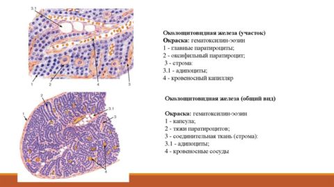
Separate trabeculae are built from two types of parathyrocytes - active cells of the parathyroid glands:
- Basophilic or main.
- Oxyphilic.
In turn, the main parathyrocytes are subdivided into two more types, differing from each other in their functional state:
- Dark (active).
- Light (low active).
The main active components of the parathyroid glands are dark basophilic parathyrocytes. They actively function, providing the functions of the parathyroid gland, due to the presence of more developed Golgi complexes and granular endoplasmic reticulums.
In the cytoplasm of dark basophilic cells there are many secretory granules, the diameter of which is not more than 400 nm, they contain parathyrin, the hormone of this endocrine organ. The parathyroid gland regulates with its help the content of calcium ions in the blood.
At the same time, the secretion of biologically active substances is carried out on the basis of the feedback principle - as soon as the calcium content in the peripheral blood falls, the production of parathyrin increases and, conversely, when the concentration of this microelement begins to exceed the norm, iron reduces the release of the hormone.
Function of glandulae parathyroideae
The parathyroid glands are responsible for controlling the exchange of calcium and phosphorus in the human body and carry it out by producing a specific biologically active substance - parathormone. The activity of the musculoskeletal and nervous systems of the human body depends on their well-coordinated work.
Attention! With the complete removal of these endocrine organs, if the doctor does not prescribe a full replacement hormone therapy, as required by the instructions, the patient may die against the background of tetany.
The principle of action of parathyroid hormone
As soon as the content of calcium ions in the peripheral blood decreases to a certain level, the receptors that are sensitive to them give the command and the parathyroid gland produces parathyroid hormone in an enhanced mode. This biologically active substance, entering the bloodstream, activates osteoclasts that extract calcium from the bones.
Thanks to this process, the blood is enriched with a much-needed trace element, however, bone loses its rigidity, and with a prolonged violation of the exchange of calcium and phosphorus, it can even be deformed. In addition, parathyroid hormone regulates the exchange of ions accountable to it, influencing the functioning of the intestines and kidneys.
In particular, under the influence of this biologically active substance, the formation of vitamin D (which enhances the absorption of calcium ions) in the intestinal lumen is activated.
Interesting! During the day, the calcium content in the peripheral blood varies significantly - it is activated during the day, along with an increase in the rate of metabolic processes, and is inhibited at night.
Therefore, the main role of the parathyroid gland in the body is the synthesis of parathyroid hormone, which catalyzes calcium metabolism.
Problems with this department endocrine system do not happen often, but their price is very high, because they can cause manifestations:
- Osteoporosis.
- Pathological fragility of bones.
- Urolithiasis.
- Cardiovascular pathologies.

But the fetus is most at risk, because the parathyroid gland, whose role in the body of a pregnant woman is so great, failing to cope with its function, can provoke the development of severe congenital pathologies in the unborn child. And only an urgent visit to an endocrinologist is the key to a return to a healthy life.
Glands and endocrine system
The endocrine system and external secretion glands perform a variety of functions in the body: they are responsible for the production of sweat, regulate metabolism, help digest food, and control the reproductive system.
The body has several types of glands - organs that produce and secrete special substances that control many life processes.
Types of glands, or what are the glands
Glands are divided into three main groups, depending on where they secrete the produced substances.
The endocrine glands are dispersed throughout the body and secrete substances - secrets - directly into the blood, which delivers them to certain organs. Endocrine glands include the hypothalamus, pituitary, thyroid, parathyroids, adrenals, pancreatic islets, testes, and ovaries - together they form the endocrine system. During pregnancy, the placenta (in addition to its other functions) also acts as an endocrine gland.
External secretion glands include those that secrete substances - excreta - onto the surface of the body (sweat, salivary, mammary, lacrimal glands) or use large ducts for this (liver, kidneys, part of the pancreas). In this group, apocrine glands are distinguished separately - large sweat and mammary glands. Sweat apocrine glands are concentrated mainly in the armpits, as well as in the lower abdomen and on the genitals.
And finally, the glands of the lymphatic system (it is more correct to call them lymph nodes). They produce special blood cells - lymphocytes - as well as antibodies to fight infection.
Lymph nodes are an integral part of the immune (defense) system of the body and increase in inflammation. The thymus gland at the base of the neck - the largest lymphoid organ in children - is more active than all other body tissues in producing lymphatic cells that destroy harmful microorganisms.
Answers from an endocrinologist to some questions from readers
I often experience discomfort due to the strong smell of sweat coming from under my arms. Can I remove sweat glands?
Sweat produced under the armpits has an unpleasant pungent odor due to bacteria and yeast that break it down. To deal with this is simple: wash your armpits more often and use a good deodorant. Talk to your doctor - he can recommend you other ways to fix this problem.
If any of the main glands ceases to perform its functions, can a donor one be transplanted instead?
No, but there are other ways of treatment. If the endocrine glands stop functioning, you can take hormonal preparations. When the pancreas stops producing insulin, diabetes develops. In this case, insulin is injected.
My aunt complains of constant fatigue. Maybe she has something with the glands?
Unlikely. An underactive thyroid can cause fatigue, but other symptoms also appear. Advise your aunt to see an endocrinologist who will examine her thyroid and other glands.
When I was sick with infectious mononucleosis, a friend said that it was a disease of the glands. Is she right?
Despite the fact that one of the most characteristic signs of mononucleosis is an increase in lymph nodes, this infectious disease, caused by the Epstein-Barr virus, affects the entire body, and not just the lymph nodes.
When my son's tonsillitis gets worse, his The lymph nodes neck. Why?
The tonsils, the inflammation of which leads to tonsillitis, are part of the lymphatic defense system. They communicate with cervical lymph nodes, which swell in response to infection. If your son often suffers from tonsillitis or his glands are constantly enlarged, consult a doctor.
The doctrine of the organs of internal secretion (Endocrinologia)
To the organs of internal secretion belong special glandular organs, different in their origin, shape and structure. Topographically, they are not interconnected, they have different sizes and weights - from a few tens of grams to barely noticeable inclusions in other tissues and organs. However, their role in the body is exceptionally great. For example, the removal in young people of only one very small gland the size of a pea - the pituitary gland - leads to growth arrest, the extinction of sexual function in adults, etc., therefore, these organs have a vital importance for the body. They produce and secrete into the blood and lymph special chemicals - hormones *, which are carried with the blood throughout the body and in negligible amounts can have a strong effect on various organs and systems, stimulating or inhibiting their activity.
* (From the Greek word "gormao" - excite, set in motion.)
The endocrine glands are of primary importance in the system of so-called humoral regulation. Humoral regulation is one of the mechanisms for coordinating functions between individual cells, organs, and physiological systems. Humoral regulation is carried out by means of substances secreted by special glands, cells, tissues in the process of their metabolism. They enter first into the tissue fluid, then into the blood and are thus carried throughout the body, exerting their effect on other organs and systems.
The interaction between organs carried out by these substances occurs in close interaction with the activity of the nervous system, since their release, entry into the blood, their transfer by blood and action on other organs are carried out by a reflex.
Thus, humoral regulation, together with the nervous one, constitutes a single system of neurohumoral regulation with the leading role of the nervous system. An example of the humoral influence of metabolic products regulated by the nervous system, i.e., by a reflex, are changes in the frequency and depth of breathing during increased physical work: an increased release of carbon dioxide occurs as a result of an increased exchange regulated by the nervous system; carbon dioxide, in turn, stimulates the nervous mechanisms of the respiratory act. The function of the endocrine glands (adrenal, thyroid, sex and other glands) depends entirely on nerve impulses and is a mechanism of conditioned reflex regulation. With insufficient intake of fluid into the body, irritation of special receptors in the tissues reflexively causes the release of the so-called pituitary gland. antidiuretic hormone, which enters through the blood into the renal tissue, causing a delay in the release of water.
Each endocrine gland consists mostly of a glandular epithelial tissue, has a dense network of blood vessels and is supplied with a large number of nerve fibers from the autonomic nervous system. common feature of all these glands is the absence of excretory ducts.
All endocrine organs are functionally interconnected. Their hormones are produced and exert their physiological effect under the direct influence of the central nervous system, participating in the regulation of vital body functions.
The endocrine organs include: the pituitary gland, the pineal gland, the thyroid gland, the parathyroid glands, the adrenal glands, the pancreatic islets, the intrasecretory part of the sex glands, and the thymus gland.
Pituitary(hypophysis cerebri). The pituitary gland (Fig. 344, 345, 346), or the lower appendage of the brain, is an egg-shaped body about 1 cm in diameter, suspended on a short and thin stalk or stalk to the lower surface of the brain in the region of the gray tubercle (hypothalamus). The pituitary stalk is part of the bottom of the third ventricle, extended into a funnel (infundibulum), in front it is covered by a cross optic nerves. This small piece of iron is located in the deepening of the Turkish saddle of the main bone. Its average weight in an adult varies between 0.6 and 0.8 g.
Rice. 346. Scheme of blood circulation in the pituitary gland: a - arteries; s - sinusoidal extensions of capillaries; k - ordinary capillaries; k g - capillaries of the second order; v - veins; st - stem; IIIV - third ventricle; h - hypothalamus
The weight and volume of the gland undergo significant changes from birth to puberty. These changes are explained by the active influence that iron has on the general somatic * development of the organism, especially on the development of the skeleton. With the onset of sexual development, the pituitary gland begins to grow rapidly, almost doubling its weight by puberty. The weight of the pituitary gland in women during pregnancy increases especially noticeably, reaching up to 1.65 g.
* (From the Greek word soma, body.)
This small grayish-reddish body consists of two main lobes: anterior and posterior, but between them there is also a third lobe (intermediate), closely fused with the posterior. The division of the pituitary gland into lobes can only be seen under a microscope. The anterior lobe is somewhat larger and denser than the posterior; the pituitary stalk is more connected to the posterior lobe.
The origin of the lobes of the pituitary gland is different: the posterior one developed from the wall of the third ventricle of the brain, the anterior and intermediate - from a special pocket-like protrusion of the epithelial wall of the pharynx, which then lost contact with the pharynx. Both rudiments are combined by connective tissue into one organ, with the brain part having the character of a neuroglia, and the pharyngeal part forming the structure of a glandular organ.
The pituitary gland is abundantly supplied with sympathetic nerve fibers from the carotid plexus and from the midbrain. In general, this gland has a very close connection with the brain.
The anterior lobe of the pituitary gland (glandular part) is also called epithelial, since it consists of strands and alveoli with a large number of epithelial cells. Connective tissue threads form the thinnest looped network, densely filled with accumulations of glandular cells.
The posterior lobe of the pituitary gland is close in structure to the nervous tissue, therefore it is also called the neurohypophysis (neural part). It consists of neuroglial tissue, i.e., of the supporting tissue of the nerve elements. The tissue of this lobe is mainly built from the finest plexus of more or less narrow fibrous loops and numerous cellular elements of glia and, apparently, nerve fibers; there are no nerve cells in this tissue.
The pituitary gland is very richly supplied with blood capillaries with a wide lumen and thin walls. Numerous arteries descend from above along the pedicle, with most of them going to the anterior lobe, and a smaller number going to the back.
The blood supply to the pituitary gland is highly variable. It receives several arteries from a. carotis interna and circulus arteriosus Willisii. In the gland, in addition to capillaries, there are also peculiar sinusoidal expansions of capillaries lined with endothelium. Part of the veins of the pituitary gland, leaving the gland, goes to the area of the gray tubercle (hypothalamus), located in the bottom of the third ventricle, and, entering the medulla, again branches into capillaries, forming a second-order capillary system; the ratios present here strongly resemble the portal circulation of the liver. These ratios naturally lead one to think that the pituitary hormones can act on the nerve elements of the hypothalamus in especially high concentration and through it on other glands and organs of the body.
The pituitary gland is present in all vertebrates. Its endocrine activity is distinguished by exceptional complexity and diversity. More than 20 different hormonal substances have already been isolated from the pituitary gland - prolan, prolactin, pituitrin and many others. The following functions of the pituitary gland have been most studied: stimulation of organ formation and body weight; trophic regulation of growth processes; stimulation of the growth and development of the genital organs and puberty (stimulation of the maturation of ovarian follicles and spermatogenesis); excitation of the secretion of the mammary glands, thyroid gland, adrenal glands and other endocrine glands.
epiphysis(glandula pinealis, s. epiphisis cerebri) (see Fig. 344). The epiphysis, or pineal gland, is located, like the pituitary gland, in the cranial cavity, but on the upper side of the brain stem above the quadrigemina and the back of the visual tubercles, with which it is connected by short legs. The shape of the iron resembles a miniature spruce cone, from which it got its name. The pineal gland, like the pituitary gland, is a derivative organ of the brain - it is an outgrowth of the upper wall of the third ventricle.
In an adult, the maximum length of the gland is 10-15 mm, the diameter is 5-7 mm, and the weight is from 0.2 to 0.3 g.
The surface of the gland is smooth, and small vascular grooves stand out on it. The section of the iron is gray-red in color, the surface of the section is fine-grained, closer to the central part there are small depressions filled with irregularly shaped calcareous masses of lemon-yellow color. These are the so-called grains of brain sand (acervuli cerebri), which in the 15th century were considered "the cause of dementia." It is still debatable whether these grains of sand represent normal or pathological elements.
In its structure, the pineal gland resembles the posterior lobe of the pituitary gland (neurohypophysis). This gland should be considered as an accumulation of glial cells of the embryonic and epithelial types.
A large number of nerve fibers connect the epiphysis with the visual tubercles and with the posterior commissure of the brain; on this basis, some scientists believe that the pineal gland, being an intrasecretory organ, is otherwise, in its structure, more like a nerve center and, perhaps, functions in the same way as it does.
Very little is known about the role of the pineal gland in the body and its intrasecretory activity. The hormones of this gland have not been isolated, and this "mysterious organ" (as it was called before), which in ancient times was considered the "center of the connection between the soul and the body", has not yet been sufficiently studied. Even the great 17th-century philosopher Descartes believed pineal gland"place of the soul". But now such an idea of the role of this gland seems at least absurd and ridiculous.
Recent research is shedding some light on the role of the pineal gland in the body. Injections of extracts from this gland cause a constant increase in the amount of calcium in the blood in humans and dogs. Obviously, it must be assumed that the hormone of the pineal gland plays a role in calcium metabolism, influencing the nerve centers through the latter, which can be concluded from the fact that extracts from the pineal gland (eliglaidol) have been successfully used against epilepsy.
Observations and experiments have shown that the pineal gland inhibits the development of the reproductive system before puberty, that is, it has an inhibitory effect on the entire complex of endocrine glands, which is in charge of completing the process of sexual development.
Thus, the pineal gland in this respect acts similarly to the adrenal cortex and, as we shall see below, to the thymus gland. By the period of puberty, the influence of the pineal gland weakens. The destruction of the pineal gland in childhood leads to premature puberty. Consequently, the pineal gland has a significant impact on the human sexual sphere.
Thyroid(glandula thyreoidea). The thyroid gland (Fig. 347, 348, 349) is one of the most important endocrine glands in the system. She was given such a name for her distant resemblance to a shield. It is located on the thyroid cartilage of the larynx and the first tracheal rings and is fixed by ligaments to the lower edge of the cricoid cartilage, as a result of which it is displaced during swallowing movements of the larynx and trachea. In front and outside, the gland is almost entirely covered with muscles, therefore, under normal conditions, it is not always possible to probe it. The gland usually consists of two lobes, connected by a bridge, from which sometimes a narrow strip of the third lobe extends upward.
The volume and weight of the gland fluctuate within fairly wide limits. In addition to the age and gender of a person, its size is influenced by climate, nutrition, intoxication, infections, and many other external and internal factors that can cause changes in both the structure and nutrition of the gland. The power of the vascular network of the gland is exceptionally large. The amount of blood supplied to iron under normal conditions is enormous compared to the volume of the organ. The entire mass of the body's blood passes through this small organ about 1 time per hour.
A number of nerves approach the thyroid gland, mainly vasomotor, but some of them also have secretory functions.
In a newborn, the weight of the gland is on average 2-2.5 g, by 7 years - 6-10 g; very quickly its increase dressed in the period preceding puberty. In an adult, the normal weight of the gland reaches 30-60 g; in women it is slightly higher than in men, especially during pregnancy.
The thyroid gland is a permanent organ in all vertebrates with a similar body structure. Outside, it is covered with a smooth fibrous membrane. On the section, the tissue of the gland is dotted with small grains and dissected into separate lobules. In its structure, the gland is a connective tissue skeleton formed by partitions coming from the fibrous membrane. These partitions, or strands, intertwined with each other, form a grid, the loops of which are filled with rounded closed vesicles, lined from the inside with a single-layer cylindrical or cubic epithelium. The bubble diameter ranges from 40 to 120 μ. In connective tissue partitions containing elastic fibers, numerous arterial, venous and lymphatic vessels and nerves pass. The epithelial cells of the vesicles are in direct contact with the wide blood and lymphatic capillaries braiding the vesicles. The bubble cavity is mostly filled with a homogeneous viscous mass, insoluble in water, alcohol, ether and weak acids; this is the so-called colloid* of the thyroid gland.
* (This term should not be confused with the concept of the physical state of substances (colloidal state).)
The colloid of glandular vesicles contains up to 1% iodine, which is part of the thyroid hormone.
The thyroid gland produces, apparently, not one, but several hormones. Thyroxine, thyroglobulin, diiodothyrosine, thyroidin are currently known. The main functions of the thyroid gland are reduced to the regulation and stimulation of the growth and development of the organs of the body and the nervous system (especially the vegetative system), the stimulation of the growth and development of the sex glands, and the regulation of the general metabolism in the body. The thyroid gland also has a beneficial effect on the development of the skeleton, skin and its appendages, and in humans, on the development of mental abilities.
Parathyroid glands(glandulae parathyreoideae). The parathyroid glands are directly adjacent to the thyroid gland (see Fig. 348), located on its posterior surface, usually in the amount of four: two for each lobe of the thyroid gland. Their number can sometimes reach up to 7-8. Parathyroid glands, or, in other words, epithelial bodies, are very small, averaging 6 × 3.5 mm in size, oval-shaped formations. The average weight of all these glands together in a person is from 0.05 to 0.09 g. They are in very close connection with the thyroid gland, located either in its capsule, or even in the gland itself. In appearance, they can be compared with lymphatic nodules, only their surface is dotted with the finest network of small venous vessels. They are supplied with branches from the lower and upper thyroid arteries and have a common lymphatic system with the thyroid gland.
The parathyroid glands are covered with a very thin fibrous membrane. Connective tissue strands run parallel to the surface, and between them are clusters of glandular epithelial cells, the mass of which is penetrated by numerous wide blood capillaries. With age, the connective tissue strands noticeably thicken and then the epithelial cells unite into lobules.
The existence of the parathyroid glands was not known at all for a very long time. Due to their small size, they are difficult to find, so no one paid any attention to them, and they remained unexplored for a long time. Previously, during operations to remove the thyroid gland, the parathyroid glands were also imperceptibly removed, which always led to death with severe convulsions, but the role of the parathyroid glands was not taken into account at all.
The function of the parathyroid glands is not yet fully understood. It mainly consists in the regulation of calcium and phosphate metabolism in the blood and tissues, as well as in the ability to quickly mobilize calcium and phosphorus ions from their reserves and direct them into the blood to maintain a constant level of calcium and phosphorus in the blood. The hormone parathyroid hormone, or parathyroidin, has been isolated.
Thymus(glandula thymus). The thymus, or goiter, gland (Fig. 350) is a lymphoid-epithelial formation. It is located in the chest cavity upper section anterior mediastinum, behind the handle and body of the sternum, adjacent to the anterior surface of the trachea, to the aorta and heart bag. It consists of two mostly unequal and asymmetrical shares. Together, these lobes form an irregularly shaped pyramid, directed downwards with its concave base and upwards with its top. The top is, as it were, split at the top, and the ends of both lobes diverge like a fork (hence its name). The gland is highly developed in young animals.
The entire gland is covered with a thin fibrous capsule, from which layers extend inside, dividing the parenchyma of the gland into numerous lobules. In each lobule, a looser cortical substance is distinguished, which, without a sharp border, passes into a denser medulla.
The average weight of the gland in a newborn is approximately 13-25 g. In the future, it grows and by the beginning of puberty (14-16 years) its weight reaches approximately 30-40 g, after which it begins to progressively decrease. In childhood, the gland is poor in connective tissue and rich in glandular parenchyma; starting from the period of puberty, the gland undergoes fatty degeneration, but a certain amount of glandular tissue remains in the elderly.
The arteries that feed it partially branch on the surface and, penetrating deep into, form a rich dense capillary network inside the gland.
In its structure, the thymus gland occupies a special position among other endocrine glands. It consists of a combination of epithelial and lymphoid tissue. The presence of lymphoid tissue in the gland brings it very close and connects it with the entire lymphadenoid system. For example, the lympho-epithelial structure of the gland is very similar to the tonsils.
The epithelial cells of the gland tend to hypertrophy and merge into syncytial masses. Derivatives of the epithelial elements of the gland are special round-shaped epithelial formations, known as Hassal's bodies. These bodies are formed by concentric layering of epithelial cells and, apparently, are involved in the formation of hormonal substances of the gland. Lymphoid tissue the gland is the site of the formation of lymphocytes, i.e., it also functions as a hematopoietic organ. The thymus regulates growth processes, stimulates mineral metabolism, helping to save calcium, mineral phosphorus and magnesium, fixing their deposition in the bones. In childhood, before puberty, it inhibits the maturation of the gonads.
adrenal glands(glandulae suprarenales). The adrenal glands (Fig. 351) are located above the upper poles of both kidneys. Each of them is a small flattened, triangular formation weighing from 10 to 15 g. The outer lateral edges of the glands are rounded, and lower surfaces concave, they are adjacent to the upper convex pole of the kidney. The inner edge of the right adrenal gland is closely adjacent to the wall of the inferior vena cava, and the inner edge of the left adrenal gland is 0.5 cm away from the abdominal aorta. Between the two inner edges of the adrenal glands in front of the aorta is the celiac (solar) plexus, connected to both glands by numerous nerve fibers. Each gland is covered on the outside by a more or less dense fatty capsule, which, with its connective tissue stroma, continues into the kidney capsule.
The adrenal gland consists of two glandular parts of different origin and unequal physiological action. On the section, we will see a more powerful outer layer - a cortical, pale yellow color, rich in lipoids, developed from the epithelium of the embryonic cavity (mesoderm), and a thinner and looser inner layer - a cerebral, brownish-gray color, developed from one rudiment with a sympathetic nervous system (from the ectoderm). The cortex and medulla have nothing in common in their structure, origin, and especially in their physiological significance, so that an anatomically single organ - the adrenal gland - is two independent endocrine glands.
The adrenal gland is richly supplied with blood. It is fed by three arteries: a branch from renal artery, an artery coming directly from the aorta, and a branch from the inferior phrenic artery. thick capillary networks glands come together central vein. The vein from the left adrenal gland flows into the renal vein, and from the right - directly into the vena cava. In terms of specific blood supply, the adrenal gland is ahead of all other tissues of the body. Up to 7 ml of blood per 1 g of weight per minute passes through the vessels of the gland.
Numerous nerve fibers enter each adrenal gland from the celiac nerve (n. splanchnicus), from the celiac plexus, from the renal plexus and from the vagus nerve. A huge number of nerve endings are scattered inside the gland. Some fibers, thin and short, terminate in small swellings in the cortical substance, other fibers, thicker and longer, form a rich network in the medulla. Here the nervous network is so dense that it wraps around almost every cell of the brain tissue. No endocrine tissue has as many nerve fibers as the cells of the adrenal medulla.
The adrenal cortex consists of epithelial polygonal cells tightly adjacent to each other, containing droplets of a fat-like substance and lipoids. In addition, the adrenal cortex is rich in sulfur-containing organic substances; the most famous of these compounds are cysteine and glutathione, which, apparently, are also synthesized here.
The function of the adrenal glands has not yet been studied enough, but it is clear that it is vital for the body. Complete removal of the adrenal glands leads to inevitable death. The adrenal cortex produces many chemically active substances, but cortin and corticosterone are the most studied. These hormones affect many functions of the body - the metabolism of carbohydrates and fats, salts and water, increase working capacity and reduce fatigue, facilitate muscle activity, weaken the action of toxins and poisons, and affect the activity of the sex glands.
The adrenal medulla secretes the well-studied hormone adrenaline, which acts on the cardiovascular system, mainly on smooth muscle vessels (arteries), maintaining its tone and general blood pressure; it helps to increase the efficiency of skeletal muscles, participates in carbohydrate metabolism, converting liver glycogen into glucose, which enters the bloodstream.
Pancreas(pancreas) * . Studies of the pancreas (Fig. 352) under a microscope show that among the glandular lobules that have excretory ducts, special cell clusters in the form of islets are scattered throughout the gland, which are completely unrelated to the excretory ducts of the gland. They have a spherical shape, their sizes range from 40 to 400 μ. These islets, consisting of glandular cells, were described as early as 1869 by Langerhans and are called the islets of Langerhans; the total amount of insular tissue in humans is 1-3% or 1/35 of the weight of the pancreas. Each islet is surrounded by a dense network of blood and lymphatic vessels, which also permeate the entire cell mass of the islet. The islets are supplied with numerous branches of the vagus nerve coming here from the celiac plexus. The islets of Langerhans at first did not attach any importance to the endocrine organ at all; they were even taken for lymphatic follicles, and only later was the epithelial glandular nature of the islets established. In 1898, AI Yarotsky was the first to establish that these islets are independent organs of internal secretion.
* (From Greek: pan - whole and creas - meat, that is, a fleshy gland.)
The islet part of the pancreas produces the hormone insulin, which is very important for the body, which is involved in the regulation of carbohydrate and water metabolism in the body. It acts in the opposite way to adrenaline. In the absence or insufficiency of insulin in the body develops serious disease- Diabetes (sugar disease).
* (From the Latin word insula - island.)
gonads(glandulae sexuales). Sex glands - testicles (Fig. 353) in men and ovaries (Fig. 354) in women - have a dual service in the body. They produce germ cells necessary for reproduction, and at the same time they are the site of the formation of sex hormones that enter directly into the blood and play an extremely important role in the life of the body. Hormones - androster he and testosterone - are isolated from the testicle, folliculin and progesterone are isolated from the ovaries.
The structure of the sex glands is described above. Sex-specific intrasecretory function is possessed by interstitial tissue, consisting of cells that make up the connective tissue between the seminiferous tubules in the testicle and between the follicles in the ovary.
The interstitial tissue of the ovary is sometimes also called the gland of maturity (pubertal gland), since during puberty the most intensive reproduction of its tissue cells occurs and the influence on the formation of sexual characteristics is most clearly expressed.
The function of the interstitial tissue of the testicles is complex and diverse: it stimulates the development and growth of secondary sexual characteristics, excites the sexual instinct, participates in the regulation of carbohydrate and basal metabolism in the body, stimulates the activity of the nervous system, having a beneficial effect on the excitability and speed of mental processes.
The intrasecretory function of the ovaries belongs to the cells that make up the shell of the egg-containing (graafian) vesicle and the interstitial cells of the connective tissue of the ovary, as well as the cells of the periodically appearing corpus luteum of the ovary.
Ovarian hormones also determine sexual characteristics, participate in the regulation of metabolism, excite the sexual instinct, as well as the instinct of motherhood, prepare the female reproductive apparatus for the normal development of the fetus and the birth of a child.
glands - organs of animals and humans that produce and release special substances involved in life processes. External secretion glands (sweat, sebaceous, lacrimal, mammary, wax glands in insects) secrete their products - secrets - onto the surface of the body through the excretory ducts.
Endocrine glands do not have excretory ducts, and the hormones produced are secreted into the blood or lymph.
Some glands (kidneys, sweat glands, partly lacrimal) selectively absorb the end products of metabolism from the blood, concentrate them and excrete them, thereby preventing poisoning of the body.
Thyroid. The hormone thyroxine regulates metabolism and protein synthesis. Lack of itleadsto the disease - mexidem. The tissues become loose, the person becomes lethargic, loses appetite, and general swelling is observed. Lack of hormones in a child leads to stunting and dementia. Thyroid dysfunction may result from a lack of drinking water iodine, necessary for the synthesis of thyroid hormones. Most often this happens in mountainous areas, where melt water is not saturated with iodine.
adrenal glands. The hormones of the adrenal cortex maintain high level performance of muscle tissue, contribute to the rapid recovery of strength after tiring physical work, regulate water-salt metabolism in the body. Preparations of the adrenal cortex (cortisone) are used in the treatment of certain metabolic diseases. Removal of the adrenal cortex results in death.
The adrenal medulla secretes adrenaline. The hormones norepinephrine and adrenaline regulate the cardiovascular system, dilate the bronchi, accelerate the breakdown of glycogen in the liver, and regulate the functioning of the muscles.
Pituitary. This gland produces a lot of hormones. The anterior pituitary gland produces growth hormone, which regulates the growth and development of the body.
The lack of this hormone leads to dwarfism. Excess hormones - to gigantism.
The hormone prolactin affects the production of milk in the mammary glands.
In the anterior lobe, several hormones are formed that selectively act on other endocrine glands.
The intermediate lobe of the pituitary gland secretes a melanophoric hormone that regulates the color of the skin.
The hormone of the posterior pituitary gland - antidiuretic - regulates water-salt metabolism.
Hypothalamus - a special part of the diencephalon.
The hypothalamus and pituitary gland in their activities are closely interconnected, forming a single hypothalamic-pituitary system. control of the hypothalamus internal organs It is possible due to the fact that it regulates the functions of the pituitary gland - the main endocrine gland. The work of the hypothalamic-pituitary system is based on the principle of feedback. If any endocrine gland begins to secrete too much or too little hormones, the hypothalamus detects a deviation in their work through the blood. Then, through the pituitary gland, it regulates and restores the normal functioning of the gland.
pancreas gland through the hormone insulin affects carbohydrate metabolism. With a lack of the hormone insulin, a disease occurs - diabetes mellitus. Insulin is a protein substance, it is isolated and synthesized artificially.
Sexual glands through the hormones progesterone and androsterone regulate the formation of the body, metabolism and sexual behavior of a person. This effect is especially evident when the gonads are removed (castration) or sex hormones are introduced into the body.
Sex glands are mixed, produce several hormones and sex cells.
II. Exocrine glands
Sweat, salivary, lacrimal glands of the stomach and intestines. The pancreas is a gland of external and internal secretion.
Founder - N. Zasimova
Energy Secret -
elimination of the root cause and normalization of the functions of the endocrine system.
Purpose of Energy: l treatment of all organs of the endocrine system, normalization of all functions of the endocrine glands, normalization of hormone production, normalization and harmonization of everything related to the work of the endocrine system. Healing is in all bodies, at all levels, in all dimensions, beyond time and space. Elimination of the causes of disruption of the ES operation, correction of the consequences. Activation of all chakras. You can work both with the entire system and specifically with individual glands.
The setting has three steps.
1st step energy tuning for the treatment of the endocrine system. Self-treatment.
Stage 2 - energy boost. Treatment of other people. (The presence of the 2nd degree of Reiki is mandatory)
3rd step - Master. Energy amplification, the right to transfer settings. (The presence of the 3rd degree of the rail is mandatory)

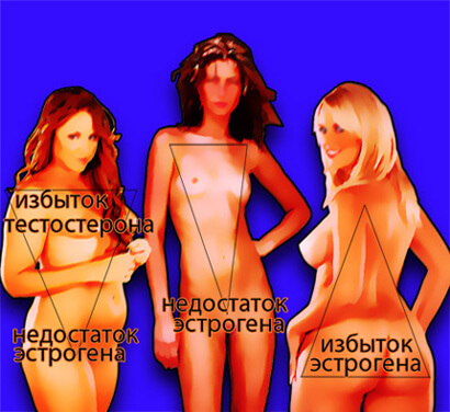
From year to year hormonal disbalance"younger". Some 10 years ago, problems with ES began after 40-45. Today, people at the age of 20-25 begin to suffer from hormonal disorders. And, unfortunately, young children are getting sick more and more often.
There are many reasons for the dysfunction of the ES: genetics, ecology, viral diseases, injury and so on. One of the main reasons is still at the energy level.
Meanwhile, it is important to maintain the smooth operation of the ES, because its violation leads to serious consequences - diabetes, infertility, gynecological problems, early menopause, malignant tumors etc.
The main symptoms of hormonal failure:
- Nervousness, mood swings, aggressiveness or tearfulness
- Sleep disturbances, drowsiness, lethargy, apathy
- Violation of sexual function and a sharp decrease in libido
- "Surprised look" - eyes wide open and shining
- Skin problems - acne, increased oiliness or dryness, frequent itching; with hair - dandruff, brittleness
- Decreased body temperature, periodic chills
- Cycle failure, early menopause, problems with reproductive function
- Frequent severe headaches
- visual impairment
- Change in appearance - h Facial erta coarsen, increase Frequent severe headaches
- Excessive sweating
- Thirst and dry mouth, with frequent trips to the toilet
- The appearance of stretch marks on the skin - on the abdomen, chest, thighs
- Crises - increased blood pressure
- Rapid weight loss with a large appetite
- Excessive hairiness - growth of hair all over the body
- Periodic causeless increase in temperature (37-37.5 ° C)
- Obesity (mostly upper body)
- Hand trembling (tremor)
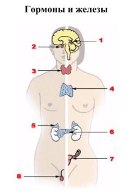
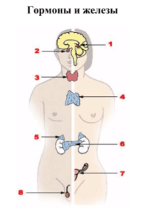
The main endocrine glands (a man is schematically represented on the left, a woman on the right):
1. Epiphysis
2. Pituitary
3. Thyroid
4. Thymus
5. Adrenal gland
6. Pancreas
7. Ovary
8. Testicle
human endocrine system —system of endocrine glands located in the central nervous system, various bodies and fabrics; one of the main control systems of the body. The regulatory influence of the endocrine system is carried out through hormones, for which characterized by high biological activity (ensuring the vital processes of the body: growth, development, reproduction, adaptation, behavior).
central link endocrine system is the hypothalamus and pituitary gland.
Peripheral endocrine system - the thyroid gland, the adrenal cortex, as well as the ovaries and testicles, glands, parathyroid glands, b-cells of the pancreatic islets.
Special place The endocrine system is occupied by the hypothalamic-pituitary system. The hypothalamus, in response to nerve impulses, has a stimulating or inhibitory effect on the anterior pituitary gland. Through pituitary hormones, the hypothalamus regulates the function of peripheral endocrine glands. For example, thyroid-stimulating hormone (TSH) is stimulated by the pituitary gland, and the latter, in turn, stimulates the secretion of thyroid hormones by the thyroid gland. In this regard, it is customary to talk about single functional systems: hypothalamus - pituitary gland - thyroid gland, hypothalamus - pituitary gland - adrenal glands.
Loss of each of the components hormonal regulation from the general system violates a single chain of regulation of body functions and leads to the development of various pathological conditions.
pineal gland still remains a mystery to scientists, it is no more than a centimeter long and weighs only 100 milligrams, but it is still one of the active organs of the body. It produces only one millionth of a gram of melatonin, but it affects the entire body. She is called the "ruler of rulers" because she indirectly controls all the underlying glands: the pituitary, thyroid, thymus, adrenals, spleen, reproductive glands.
The endocrine system produces hormones- biologically active substances, which regulate growth, reproduction and many other processes in the body.
Gland is an organ that produces and releases certain substances into the body or onto the surface of the body. Endocrine glands, or endocrine glands, do not have excretory ducts and secrete hormones directly into the blood or lymph. Exocrine glands, or glands of external secretion, include salivary, sweat, and milk glands. Exocrine glands have excretory ducts through which they secrete their secret to the surface of the body or into some cavity inside the body.
The level of hormones in the blood is regulated by feedback mechanisms, i.e. with an excess of any hormone in the blood, its secretion by the gland is inhibited, and with a deficiency, on the contrary, it is activated.
For example, thyroxine (thyroid hormone) stimulates metabolic processes: if there is too much of it, the metabolism speeds up, if it slows down a little. Deficiency of thyroxine forces the pituitary gland to secrete thyroid-stimulating hormone in order to force the thyroid gland to produce thyroxine. An excess of thyroxin has the opposite effect.
Violation of the natural hormonal regulation leads to endocrine and other diseases.
glands.
In humans, glands include:
1.adrenal glands located near the upper poles of the kidneys, which produce, among others, the stress hormone adrenaline;
2. pancreas;
3. six salivary glands- two parotid, two submandibular and two sublingual;
4. thyroid and four parathyroid;
5. pituitary and epiphysis(in the brain);
6. two lacrimal glands and, finally, two dairy, which produce milk in women and are atrophied in men.
Among women In addition, there are two ovaries in which eggs mature, two Bartholin glands, and two skene glands that provide vaginal lubrication.
In men there is a prostate gland, two cooper glands, whose role is in the formation of sperm, and seminal vesicles - "factories" of spermatozoa. Thus, men have a total of 27 glands, while women have 28.
The total weight of the glands is 3 kg, and their volume is the size of a soccer ball.
The endocrine system is made up of glands that produce hormones.
 Together with the nervous system, it regulates and coordinates the entire vital activity of the body. Along with growth and reproduction, in which it plays a key role, the endocrine system regulates many other processes. However, the nervous and endocrine systems act differently. In the nervous system, signals are transmitted in the form of electrical impulses. The endocrine system releases chemical messengers, hormones, into the blood. Under the control of hormones, all stages of the development and life of the body proceed - from inception to
old age. Blood delivers hormones to their destination, where these biologically active substances act on cells, accelerating or slowing down the processes occurring in them. Unlike nerve impulses, hormones act slowly and cause a long-term effect.
Pituitary controls the activity of most other glands. The pituitary gland itself is under the control of the hypothalamus - a small brain structure formed by a cluster of nerve cells.
Hypothalamus provides a direct link between the endocrine and nervous systems.
Glands of the endocrine system located in the head chest and abdominal cavity. Chief among them are the pituitary, thyroid, parathyroid, and adrenal glands. The pituitary gland, which produces more than 9 hormones, regulates the activity of most other endocrine glands and is itself under the control of the hypothalamus.
The thyroid gland regulates growth, development, intensity of metabolism in the body. Together with the parathyroid gland, it also regulates the level of calcium in the blood.
adrenal glands also affect the intensity of metabolism and help the body resist stress.
Pancreas regulates blood sugar levels and at the same time acts as an external secretion gland - secretes digestive enzymes through the ducts into the intestines.
endocrine sex glands- testes in men and ovaries in women - combine the production of sex hormones with non-endocrine functions: germ cells also mature in them.
Pituitary
This tiny gland located at the base of the brain, which produces more than 9 hormones, regulates the activity of the endocrine system. Some of its hormones directly control certain functions, for example, growth hormone stimulates growth. Others act on individual endocrine glands, forcing them to produce their own hormones. So, the pituitary follicle-stimulating hormone enhances the synthesis of the female sex hormone estrogen in the ovaries.
The pituitary gland is made up of 2 lobes. The larger anterior produces and secretes most of the pituitary hormones. Their secretion is stimulated by hormones of the hypothalamus (diencephalon). The posterior (smaller) lobe of the pituitary gland stores and releases 2 hormones synthesized in the hypothalamus.
adrenal glands
The adrenal glands are located on the kidneys, covering them from above in the form of a cap. Each adrenal gland consists of a cortical layer and a medulla. The bark secretes corticosteroid hormones. They regulate metabolic processes and the concentration of certain substances in the blood. The medulla secretes adrenaline. When the brain perceives the situation as
dangerous or stressful, it sends a signal to the adrenal glands, causing them to secrete adrenaline. Thrill-seekers riding a rollercoaster experience the hormone adrenaline.
Adrenaline, secreted by the adrenal glands, helps the body resist danger. It makes the heart beat faster, speeds up breathing and increases blood flow to the muscles. The release of adrenaline into the blood is the preparation of the body to face danger.
to face or, conversely, flee from it.
Some glands regulate and maintain the normal functioning of the body by sending signals through the blood vessels that permeate our body.
The signals sent by these glands are special substances - hormones that are released into the blood and with it reach the organs where they have their effect. So, hormones are like encrypted signals sent by individual points of the body.
to other points where there is a key to decrypt them; these substances in the smallest doses can stimulate some processes, slow down others and give rise to new ones.
Organ and main hormones:
Hypothalamus
Oxytocin, Vasopressin
Pituitary
Thyroid Stimulating Hormone (TG), Gonadotropic Hormone (FSH), Luteinizing Hormone (LH), Adrenocorticotropic Hormone (ACTH), Prolactin, Growth Hormone (GH) (Somatotropin), Melanostimulating Hormone (MSH)
Thyroid
Thyroxine, Triiodothyronine, Calcitonin
Parathyroid
Parathormone
Pancreas
insulin, glucagon
Gastrointestinal hormones
Gastrin, Enterogastron, Pancreozymin, Secretin, Cholecystokinin, Enterocrinin
adrenal glands
Cortisol, Aldosterone, Adrenaline, Norepinephrine
gonads
Estrogens, Testosterone, Estradiol, Progesterone
Be healthy!
DO NOT allow the appearance or intensification of the disease!
Start Your Treatment NOW!
You can receive the setting in person or remotely. RECORD FOR SETTING- registration
We agree on the date and time of the setup, I send you all the necessary materials for work.